| Starting
material |
Human iPSC line |
| Donor |
Caucasian adult male (skin
fibroblast), Genotype APOE 3/3 |
| Format |
Cryopreserved cells |
| Differentiation
method |
opti-ox deterministic programming |
| Vial size |
Small: >1.5 x 10^6 viable cells |
| Recommended
seeding density |
37,000 to 39,500 cells/cm2 |
| Seeding
compatibility |
6, 12, 24, 48, 96 & 384 well
plates |
| Quality
control |
Sterility, protein expression (ICC),
functional phagocytosis, GFP expression (flow cytometry) |
프로토콜
GFP ioMicroglia는 냉동 보존 형식으로 제공되며 권장 배지에서 재생 시 빠르게 성숙되도록 프로그래밍되어 있습니다.
1) 유도단계 : bit.bio에서 Induction 후 제공
2) Phase 1 : 고객이 세포를 수령 후 plating 하여 안정화시키는 기간 (1일)
3) Phase 2 : 세포가 성숙하는 기간 (9일)
4) Phase 3 : 세포를 유지하는 기간 (1일)
세포는 10일째 되는 날부터 실험에 사용할 수 있습니다.

참고 데이터
1. Flow cytometry analysis of GFP expression at day 11 and day 21 for GFP ioMicroglia
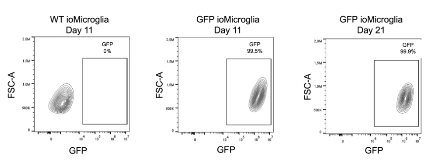
Flow cytometry analysis demonstrating GFP expression in over 99.5% of cells for GFP ioMicroglia cultured until day 11, with no GFP expression seen in ioMicroglia Male (io1021). At day 21, the percentage of cells expressing GFP and the intensity does not decrease over time, indicating there is no silencing of the reporter gene.
2. GFP ioMicroglia express key microglia markers
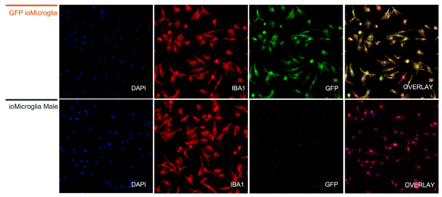
Immunofluorescent staining on day 10 post-revival demonstrates similar homogenous expression of microglia markers P2RY12 and IBA1 and ramified morphology in GFP ioMicroglia compared to ioMicroglia Male (io1021). GFP expression can be visualised throughout the cytosol in every cell for GFP ioMicroglia, but not in the WT control. 10X magnification.
3. GFP ioMicroglia show ramified morphology by day 10
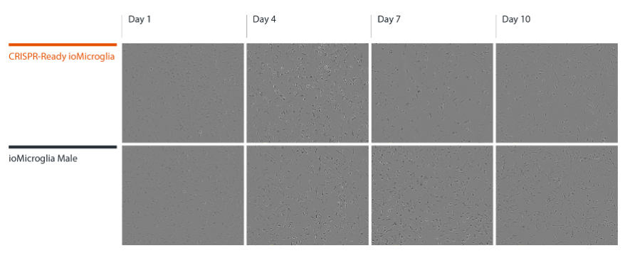
GFP ioMicroglia mature rapidly, and key ramified morphology can be identified by day 4 and continues through to day 10, similarly to ioMicroglia Male (io1021). Day 1 to 10 post-thawing; 100x magnification.
4. GFP ioMicroglia secrete pro-inflammatory cytokines upon activation
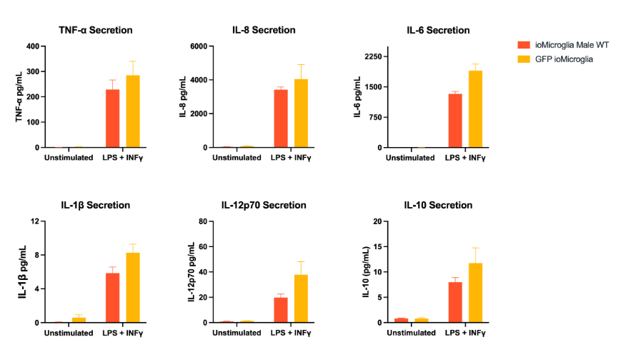
GFP ioMicroglia were stimulated at day 10 post-thaw with LPS 100 ng/ml and IFNɣ 20 ng/ml for 24 hours. Supernatants were harvested and analysed using MSD V-plex Proinflammatory Kit. GFP ioMicroglia secrete TNF⍺, IL-6, IL-8, IL-1b, IL-12p70 and IL-10 in response to the inflammatory stimuli. GFP ioMicroglia demonstrate a similar response to ioMicroglia Male (io1021), as expected. Three technical replicates were performed per lot.
5. Phagocytosis of E. coli particles by GFP ioMicroglia
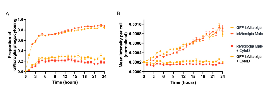
(A) Phagocytosis assay using pHrodo™ E. coli BioParticles™ at day 10 post-thaw demonstrates efficient uptake of bacteria particles by GFP ioMicroglia in comparison to ioMicroglia Male (io1021) +/- cytochalasin D control (an inhibitor of actin polymerisation). The graph displays that the proportion of cells phagocytosing E.coli particles over 24 hours for three technical replicates.
(B) The graph displays that the degree of cells phagocytosing E.coli particles over 24 hours. Images were acquired every 30 mins on the Incucyte® looking at red fluorescence and phase contrast. Three technical replicates were performed.
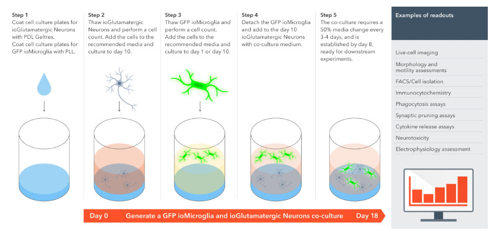
6. Easy-to-use co-culture protocol for GFP ioMicroglia with ioGlutamatergic Neurons
GFP ioMicroglia를 ioGlutamatergic Neurons 및 관련 질환 모델과 함께 공동 배양하는 방법은 다음과 같습니다.
